What weights are given to MRI images and why do we care?
Not to be confused with the featured image, which depicts 3 brains wherein similar semantic concepts, though varied between the individuals, occur in similar parts of their brains, the black and white image below shows a single brain, weighted differently in the fMRI process.
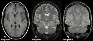
Image: Magnetic Resonance Imaging may weight the images to obtain different views of the same region. The above image shows the same region of the brain, weighted 3 ways: with longitudinal relaxation time (T1), transverse relaxation time (T2) and proton density (PD).
First, to explain what is being measured by T1 and T2:
Multiple transmitted radiofrequency pulses can be used in sequence to emphasise particular tissues or abnormalities. A different emphasis occurs because different tissues relax at different rates when the transmitted radiofrequency pulse is switched off. The time taken for the protons to fully relax is measured in two ways. The first is the time taken for the magnetic vector to return to its resting state and the second is the time needed for the axial spin to return to its resting state. The first is called T1 relaxation, the second is called T2 relaxation.
Source: National Institutes of Health website
So What is this miracle diagnostic tool called Magnetic Resonance Imaging (MRI)?
In general, the operation of an MRI depends on the state of fluid retention within the subject’s body, which may be detected by Nuclear Magnetic Resonance. “Nuclear” refers to the type of atom targeted by the MRI, most often that being hydrogen.
Nuclear magnetic resonance (NMR) is a physical phenomenon in which nuclei [most commonly, hydrogen nuclei] in a magnetic field absorb and re-emit electromagnetic radiation. This energy is at a specific resonance frequency which depends on the strength of the magnetic field and the magnetic properties of the isotope of the atoms.
Source: Wikipedia
According to the U. S. National Library of Medicine, National Institutes of Health:
Magnetic resonance imaging (MRI) uses the body’s natural magnetic properties to produce detailed images from any part of the body. For imaging purposes the hydrogen nucleus (a single proton) is used because of its abundance in water and fat.
The hydrogen proton can be likened to the planet earth, spinning on its axis, with a north-south pole. In this respect it behaves like a small bar magnet. Under normal circumstances, these hydrogen proton “bar magnets” spin in the body with their axes randomly aligned.
When the body is placed in a strong magnetic field, such as an MRI scanner, the protons’ axes all line up. This uniform alignment creates a magnetic vector oriented along the axis of the MRI scanner. MRI scanners come in different field strengths, usually between 0.5 and 1.5 tesla.
. . . .
There are no known biological hazards of MRI because, unlike x ray and computed tomography, MRI uses radiation in the radiofrequency range which is found all around us and does not damage tissue as it passes through.
The ncbi.nlm.nih.gov website referenced by the link above describes that an MRI detects hydrogen atoms within the body. The site explains the use of magnetic resonance and radio waves to cause the body’s hydrogen atoms to line up, and how manipulation of the magnetic and radio wave factors allow detection of different states of hydration.
Below is a brief definition of the MRI from myvmc.com. However, the above-referenced article explains that different radio waves may be employed to target specific states of the body.
MRI (magnetic resonance imaging) is a diagnostic technique used to create images of the body like a CT. It is non-invasive (It does not enter the body cavity) and requires no radiation, instead it is based on the magnetic fields of the hydrogen atoms in the 3D Magnetic Resonance Imaging (3D MRI)body. By scanning the body, MRI is able to provide computer-generated images of the body’s internal tissues and organs. MRI usually scans the body in an axial plane (ie. cutting the body into slices from front to back). Usually the images are 2-dimensional, where the MRI images are usually presented in slices from top to bottom. However, using sophisticated computer calculation, these 2-dimensional slices can be joined together to produce a 3-dimensional model of the area of interest being scanned. This is called 3D MRI.
What is missing from the above definition is the historical context, found at www.two-views.com, from which we quote a three events as follows.
A quick history of the MRI begins in 1882 with Nichola Tesla having discovered the Rotating Magnetic Field in Budapest, Hungary.
. . . .
In 1937 Columbia University Professor Isador Rabi discovered Nuclear Resonance Imaging–he recognized that the atomic nuclei show their presence by absorbing or emitting radio waves when exposed to a sufficiently strong magnetic field.1993 – Functional MR imaging of the brain is introduced.
It was not until 2000 that the MR scan became widely used as a diagnostic tool for many medical purposes. But for brain imaging, it is the functional MRI developed in 1993 that is most useful.
To read about Functional Magnetic Resonance Imaging, visit the next page.
Image source: By KieranMaher at English Wikibooks [Public domain], via Wikimedia Commons